
[ad_1]
Anderssen, K. E., Gabrielsen, G. W., Kranz, M., & Collard, F. (2022). Magnetic resonance imaging for non-invasive measurement of plastic ingestion in marine wildlife. Marine Pollution Bulletin, 185(PB), 114334. https://doi.org/10.1016/j.marpolbul.2022.114334
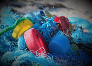
Plastics in the sea
The world has been concerned about plastics polluting the environment, and it’s right to do so. Plastics not only cause suffocation in animals, but they can also be ingested. Focusing on marine organisms, seabirds consume plastics globally, and it’s believed half of seabird species have plastic in their stomachs! For example, a study found plastics inside two-thirds of the 555 fish species they researched, and 210 of them are commercially fished.
One thing is how this affects one specific being, and another is how much it influences the whole population, which is something hard to determine, and it needs more research. The way to conduct these studies is invasive and limited, which is the reason why this particular study aims to use magnetic resonance imaging. MRI is quicker and non-invasive, so if it could be applied to plastic ingestion monitoring, it would greatly improve future studies.
For now, those invasive ways of quantifying plastics in an animal’s stomach consist of two options. One is to sacrifice the animal for dissection, which constrains how many animals can be studied, and if that particular species is endangered it can be tricky. Sadly, it often means that to protect those species, researchers have to sacrifice some of them. The second option consists of stomach flush, thrusting water down their stomachs so they regurgitate its contents. The animal is not sacrificed, but it’s highly invasive and stressful for them, and it’s not even a secure way of knowing everything that is inside their stomach.
There is always using found animal corpses, but it’s not enough for thorough studies, as it is quite hard to find enough samples, and it depends on the state they are in. Not only that, but to make an accurate research it needs random sampling which is not met when it depends on found carcasses, and high plastic consumption can lead to higher mortality, influencing the results.
Finding a way
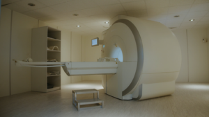
This study strives to find other ways to conduct this type of research. Sometimes, getting inspiration from other fields can be eye-opening. In this particular case, the technology they decided to investigate is more commonly used in medicine (Figure 2).
Magnetic resonance imaging (MRI) is an application of nuclear magnetic resonance (NMR), but no need to worry about its name, there’s no harmful ionizing radiation involved! It works thanks to strong radio frequency pulses (rf) to stir the system away from equilibrium to study its characteristics. Feathers and muscles are not a problem, as this method can be used inside the sample.
Not only does it provide imaging, but it also gives information on the physical and chemical properties of the materials found inside organisms. The 1H spectrum informs of the chemical properties, as the pattern and width of the lines in the spectrum provide details. On the other hand, for the physical characteristics, time is used – specifically the time it takes the system to go back to equilibrium after the rf pulse. It’s called the T2 relaxation.
But does it work?
There is a variety of techniques and equipment of NMR and MRI for different purposes, so the ones used in this case were, first, a small animal MRI imager. It carries out imaging on the samples. However, it depends on the relaxation time, as when it is too short it can’t measure properly. This is the case for plastics, so a second NMR system is necessary. This one works for materials research, and can measure with short relaxation times; however, it needs small sample sizes to perform properly.
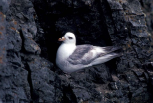
To test this method, they focused on a species of seabird, the northern fulmar (Fulmaris glacialis) (Figure 3), which is the species employed by the Arctic Monitoring and Assessment Program because of their feeding habits, which happens on the water surface. For the trials, they used four stomachs from deceased specimens, two of them artificially filled with plastics, and the other two empty. To check if this method could detect types of plastic, there were two kinds of plastic introduced. They weren’t randomly selected, as they’re the two of the most common found in seabirds: pieces of LDPE plastic bag and pipette tips of polypropylene, like the ones used in labs.
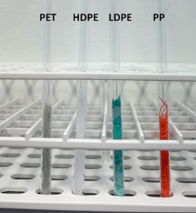
Besides the ones inside the stomachs, they also tested other different types of plastic with the NMR spectrometer, inside tubes (Figure 4). These types of plastic can be classified as PET plastics, which make drink bottles and food packaging; HDPE plastics from shampoo, detergent and cleaning bottles; and LDPE plastics from plastic bags; then we find the pipette tips and fishing rope.
The MRI tests were able to measure one type of plastic directly inside a stomach, and another one to be indirectly detected. When NMR measurements were applied, different signals came out for each plastic type, distinguishable between them. These results mean that specialized MRI sequences could be used to image different types of plastic.
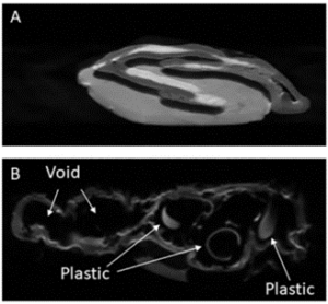
Let’s take a look inside
As we can see in Figure 5, the pipette tips are visible, but the LDPE plastic bags aren’t, looking like void spaces in the stomach. Their presence can be deduced by the way they shape the stomach, meaning that even if we can’t see the plastic, we can know it’s there. Despite this, it’s not an ideal situation, as there could be other objects with no NMR signals, like rocks, and it’s not better than having equipment that could actually measure what is necessary in these cases. As with the T2 distributions, there are consistent peaks for plastic types.
With suitable equipment, these studies could be carried out with entire birds, and also fish, turtles, and other animals. Right now, it’s not possible to take standard MRI equipment to the field because of its size and energy requirements. Some options and developments are being considered, like a single-sided NMR sensor, where the magnetic field is projected laterally into the subject, instead of placing it inside the magnet. It’s more practical for bigger samples as well as portable. They’re not commercially available yet, but the existing ones could be adapted for seabirds. Once the equipment is accessible, there needs to be a validation process to determine parameters, detection limits and calibration.
Besides needing adequate equipment, there are other issues – plastics that have been in the stomach could have some degradation. On top of that, animals also need to eat, and the signal for food needs to be untangled from plastics’ signals.
Opening the door to the future
Developing non-invasive technology to better study processes in our world would imply several benefits. As it is non-invasive, it makes it possible to study endangered species further.
Not only is it better for the animals themselves, but it would allow time and money to be saved, instead of them being spent on dissections in the lab. The time measurement depends on the system, but it’s likely to take around a minute or two.
Although it was possible to identify types of plastic and to carry on the imaging of them, this tech is far from being used in the wild. Some development needs to happen before being applied in the field.
It was possible to find plastic directly and indirectly through magnetic resonance inside stomachs. This is a step further into a future where this kind of biomonitoring is more efficient, potentially helping scientists worldwide.
I have a degree in Sea Science from the University of Barcelona, Spain. My main scientific interests are about conservation and ecology, especially anything about marine invertebrates. I find them the most fascinating creatures on Earth, strange yet so familiar. On a visit to the beach as a baby, I learned to crawl by going towards the sea at full speed! I enjoy reading, drawing, and writing fantasy novels in my spare time.
[ad_2]
Source link