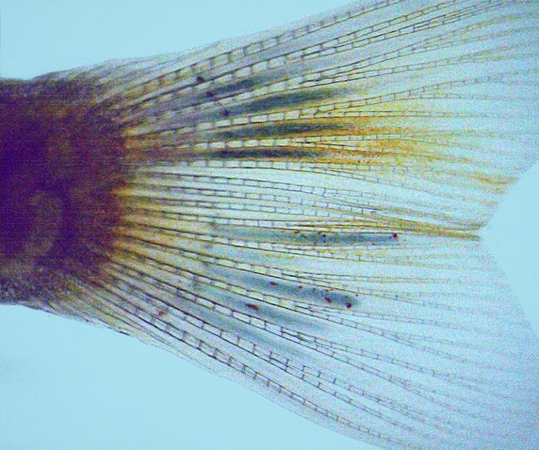
Conductive polymers (blue) formed in the tail fins of living zebrafish.Credit: X. Strakosas et al./Science
An injectable gel tested in living zebrafish can use the animals’ internal chemistry to transform into a conductive polymer.
The discovery, reported on 23 February in Science1, could lead to the development of electronic devices that can be implanted into body tissues such as the brain without causing harm.
When the gel is mixed with the recipient’s own metabolites — chemicals generated by the body’s processes — a chain reaction turns it into a solid but flexible material.
“We are performing a lot of experiments with these materials to grow electrodes and electronics around cells,” says study co-author Magnus Berggren, a materials scientist at the Linköping University in Sweden. He adds that the work could ultimately improve technologies for deep-brain stimulation, for example, or help damaged nerves to regrow.
Internal electronics
Electronic devices or circuitry that can be implanted in the body have many potential applications in medicine and research, such as helping the brain to communicate with prosthetic limbs, or even enhancing memory. But conventional electronic materials can cause inflammation or scarring, and they often deteriorate inside living tissue and eventually stop working.
The brain-reading devices helping paralysed people to move, talk and touch
Although there has been progress towards developing soft, flexible electrodes, it is difficult to get them into the body in a non-invasive way, says Berggren. If you want to insert something deep into the brain, for example, “you will basically make it cut all the way through”, he says.
Berggren’s team wanted to create a material that was conductive, but stable in the long term, non-toxic and of a consistency that allows it to be injected.
The mixture they developed contains the chemical building blocks for a conductive polymer, along with enzymes. When injected into living tissue, the gel reacts with the common metabolites glucose and lactate, which causes the gel to polymerize into a much firmer — although still soft — material. Working with a group led by chemical biologist Roger Olsson at Lund University in Sweden, the researchers used this approach to generate polymer ‘electrodes’ inside the fins and brains of living zebrafish (Danio rerio). They also used it in the nervous tissue of leeches and in muscle tissue from chickens, pigs and cows.
Because the material doesn’t polymerize until it is inside the body, and is “compliant, soft and biocompatible”, it eliminates mechanical differences between typical electrode materials and living tissue that make some medical implants so invasive, says Timir Datta-Chaudhuri, an electrical engineer at the Feinstein Institutes for Medical Research in Manhasset, New York.
Alternative approach
The idea of using a living tissue’s chemistry to create a conducting material inside the body is not new. In 2020, researchers reported engineering an enzyme to be expressed in genetically modified neurons in the nematode worm Caenorhabditis elegans. This caused the cells to produce conductive polymers2.
Restoring smell with an electronic nose
That approach could not be used in people, says Sahika Inal, a bioengineer at the King Abdullah University of Science and Technology in Thuwal, Saudi Arabia. For her, the value of the latest research is that the gel reacts with substances that the body produces naturally, and it does not require the organism to be genetically modified. “I think this technology is providing alternative thinking. Instead of changing the same device’s software, why don’t we just completely get rid of that device and make the device inside the cell?”
There are still many barriers to be overcome before the injectable substance can be tested in people. Even though the polymer is highly conductive, for example, there is currently no way to make it functional by connecting it to an outside electricity source.
The researchers also need to do more tests to establish that the approach is safe. They didn’t observe any unusual behaviour in the zebrafish after injecting the solution into their brains, but they monitored the animals for only three days after the procedure. “They need to look at long-term chronic responses,” says Inal.


